
Retinal Detachment - Procedure, Risk, Type & Treatment In India
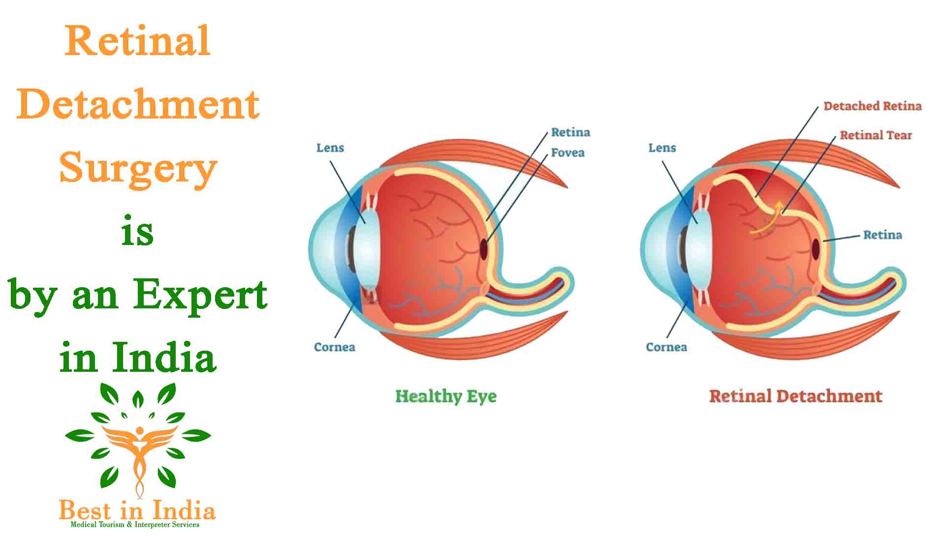
When the retina is torn from its natural location in the back of the eye, a retinal detachment ensues. The retina is a layer of light-sensitive tissue that lines the inside of the eye and transmits visual signals to the brain via the optic nerve. If left untreated, retinal detachment can result in permanent eyesight loss.
Types of Retinal Detachment
- Rhegmatogenous Retinal Detachment: A hole or crack in the retina that allows fluid to enter the gap between the retina and other layers of the eye causes a rhegmatogenous retinal detachment.
- Exudative or Secondary Retinal Detachment: Inflammation causes fluid to build up beneath the retina without a hole or break, which leads to an exudative retinal detachment.
- Tractional Retinal Detachment: As seen in cases of diabetic retinopathy, a traction retinal detachment happens when fibrovascular tissue on the surface of the retina pushes the retina away from the underlying tissues.

Symptoms of Retinal Detachment
- Most often felt in the temporal (outside, away from the nose) region of vision, flashes of light (photopsia)
- Floaters: black cobweb-like spots moving in front of the eye
- A curtain-like shadow coming in the field of vision
- Sudden Vision Loss when macula is detached
Who is at risk for Retinal Detachment?
Although a retinal detachment can happen at any age, those over 40 are more likely to experience one. These are typical risk factors for getting RD:
- High Myopia, especially more than 5 Dioptre, after cataract surgery
- Retinal detachment in the other eye
- Family history of RD
- Presence of other eye diseases such as retinoschisis, degenerative myopia, or lattice degeneration Following an eye injury
What are the treatment options for Retinal Detachment?
The outside process for treating retinal fractures or holes is cryopexy, which involves freezing the affected area. To "weld" the retina together during laser treatment, tiny burns are applied all around the hole. The area surrounding the hole is frozen during cryopexy, which aids in reattaching the retina. Scleral buckling and complicated vitreoretinal surgery with Silicon oil or gases are two surgical procedures used to treat retinal detachments.
- In scleral bucking: A silicone band is wrapped around the eyeball and attached with stitches in order to gently press the eye's wall up against the detached retina.
- In a vitrectomy: The doctor creates very small incisions in the sclera (white portion of the eye). In order to press the retina into position, the vitreous gel is removed using devices used in vitreoretinal surgery and refilled with silicone oil or gases. To seal the breaks, laser or cryopexy procedures are used.
Nearly 90% of retinal detachment cases can be effectively treated with modern vitreoretinal instrumentation. If the retinal detachment is fixed before the macula, the area of the retina responsible for fine, detailed vision, and detaches, the visual outcome will be favourable. When you experience any flashes, floaters, or a curtain in your field of vision, it's critical to get in touch with your retina surgeon as soon as possible.